CALB1 Antikörper
-
- Target Alle CALB1 Antikörper anzeigen
- CALB1 (Calbindin (CALB1))
-
Reaktivität
- Human
-
Wirt
- Maus
-
Klonalität
- Monoklonal
-
Konjugat
- Dieser CALB1 Antikörper ist unkonjugiert
-
Applikation
- Western Blotting (WB), Immunohistochemistry (IHC), Immunofluorescence (IF)
- Kreuzreaktivität
- Rind (Kuh), Human, Maus, Ratte (Rattus)
- Aufreinigung
- Protein G purified culture supernatant
- Immunogen
- Recombinant full length human calbindin purified from E. coli.
- Isotyp
- IgG2a
-
-
- Applikationshinweise
- Optimal working dilution should be determined by the investigator.
- Beschränkungen
- Nur für Forschungszwecke einsetzbar
-
- by
- Prof. Merighi, Laboratory of Neurobiology, Department of Veterinary Sciences, University of Turin
- No.
- #104494
- Datum
- 02.08.2023
- Antigen
- CALB1
- Chargennummer
- GS116G
- Validierte Anwendung
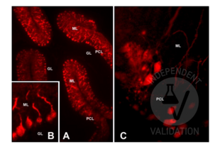
- Immunohistochemistry
- Positivkontrolle
Adult (>2 months) CD1 mouse cerebellum (6 µm glass-mounted microtome sections)
Postnatal day 6-7 CD1 mouse cerebellum (cultured cerebellar slices)
- Negativkontrolle
Control slices were processed for each experimental procedure, omitting the primary antibody; overnight incubation in diluent solution only.
- Bewertung
Passed. The CALB1 antibody ABIN6254097 works in IHC-P at a 1:50 dilution with or without tyramide amplification.
- Primärantikörper
- ABIN6254097
- Sekundärantikörper
goat anti-mouse IgG (H+L) AF488-conjugated antibody (Thermo Fisher Scientific, A11034, lot 2380031)
- Full Protocol
- Indirect IMF on microtome sections
- Perfuse adult (>2 months) CD1 mice with paraformaldehyde 4% in 0.1 M phosphate buffer pH 7.4 and post-fix in the same fixative for an additional 2 h at RT.
- Wash, dehydrate, and embed samples in paraffin wax.
- Wash several times with 0.01 M PBS.
- Cut the cerebellum with a microtome into 6 µm sections and mount them on glass slides.
- After paraffin removal, incubate sections for 1 h at RT in PBS containing 1% albumin from chicken egg white (Sigma, A5378) and 0.3% Triton-X-100 (BioRad, 161-0407, lot 00583) to block non-specific binding sites.
- Incubate sections with primary mouse anti-CALB1 antibody (antibodies online, ABIN6254097, lot GS116G) diluted 1:50, 1:100, and 1:200 in 0.1 M PBS-BSA-PLL ON at RT.
- Wash 3x 5 min in 0.01 M PBS.
- Incubate sections with secondary goat anti-mouse IgG (H+L) AF488-conjugated antibody (Fisher Scientific, A11034, lot 2380031) diluted 1:500 in 0.1 M PBS for 1 h at RT.
- or alternatively with tyramide amplification:
- Incubate sections with Poly-HRP-conjugated secondary antibody for 1 h at RT.
- Wash sections 3x 5 min in 0.01 M PBS.
- Incubate sections with Tyramide working solution (for 5 sections: 5 μL 100X Tyramide stock solution, 5 μL 100X H2O2 solution, 500 µL 1X Reaction buffer) for 10 min at RT.
- Stop the reaction with the Reaction Stop Reagent working solution.
- Wash 3x 5 min in 0.01M PBS.
- Mount specimens in Fluoroshield (Sigma-Aldrich, F6182, lot MKCB0153V).
- Acquire Images with Leica DM 6000B fluorescence microscope equipped with a digital camera at 20-40x magnification.
- Indirect IMF on cultured cerebellar slices
- Euthanize CD1 mice at postnatal day 6-7 (P6-P7) with an overdose of 60 mg⁄100 g body weight sodium pentobarbital (Merck Life Science, Y0002194).
- Remove the brain removed from the skull while the head is kept submerged in ice-cooled Gey’s solution (Sigma-Aldrich) supplemented with glucose and antioxidants (for 500 mL: 4.8 mL 50% glucose, 0.05 g ascorbic acid, 0.1 g sodium pyruvate).
- Dissect the cerebellum from the brain.
- Cut 350 μm thick parasagittal slices of the cerebellum with a McIlwain tissue chopper (Brinkmann Instruments).
- Plate two to three slices onto a Millicell Cell Culture Insert (Merck Life Science, PICM0RG50).
- Place each insert inside a 35 mm Petri dish containing 1 mL of culture medium consisting of 50 % Eagle basal medium (BME, Sigma Chemicals), 25 % horse serum (Gibco by Thermo Fisher Scientific), 25 % Hanks balanced salt solution (Sigma-Aldrich), 0.5 % glucose, 0.5 % 200 mM L-glutamine, and 1ؘ % antibiotic/antimycotic solution.
- Incubate slices at 34 °C in 5 % CO2 for up to 20 d in vitro (DIV). Change the medium twice a week. Slices were allowed to equilibrate to the in vitro conditions for at least 4-6 DIV before IMF.
- Remove the culture medium from the dish and fix the slices in 1 mL of 4 % paraformaldehyde (Merck Life Science, P6148) in PBS for 1 h.
- Wash 3x 5 min in 0.01 M PBS.
- Incubate fixed cultures in PBS containing 1 % Triton X-100 (BioRad, 161-0407, lot 00583) for 10 min.
- Wash 3x 5 min in 0.01 M PBS.
- Incubate cultures ON at 4 °C under continuous stirring in PBS containing 1 % albumin from chicken egg white (Sigma, A5378) and 0.3 % Triton-X-100 (BioRad, 161-0407, lot 00583) to block non-specific binding sites.
- Incubate cultures with the primary mouse anti-CALB1 antibody (antibodies online, ABIN6254097, lot GS116G) diluted 1:50 in PBS-BSA (Sigma, A7906)-PLL (Sigma, P1524) ON at RT.
- Wash 5 x 5min in PBS.
- Incubate cultures with the secondary anti-rabbit antibody Alexa Fluor 488 diluted (Invitrogen by Thermo Fisher Scientific, A11034, lot 2380031) 1:500 in 0.1 M PBS for 1 h at RT.
- Wash 3x 5 min in 0.01 M PBS.
- Mount specimens in Fluoroshield (Sigma-Aldrich, F6182, lot MKCB0153V).
- Acquire Images with Leica DM 6000B fluorescence microscope equipped with a digital camera at 20-40x magnification.
- Anmerkungen
For indirect IMF on cerebellum paraffin sections, antigen retrieval treatment was also tested. In this case, sections were processed for microwave antigen retrieval for 10 min (95-100 °C) in 10 mM sodium citrate buffer (pH 6.0). After 20 min of spontaneous cooling, sections were washed twice for 5 min with distilled water and twice for 5 min with PBS.
Validierung #104494 (Immunohistochemistry)![Erfolgreich validiert 'Independent Validation' Siegel]()
![Erfolgreich validiert 'Independent Validation' Siegel]() ValidierungsbilderProtokoll
ValidierungsbilderProtokoll -
- by
- Prof. Merighi, Laboratory of Neurobiology, Department of Veterinary Sciences, University of Turin
- No.
- #104530
- Datum
- 02.08.2023
- Antigen
- CALB1
- Chargennummer
- GS116G
- Validierte Anwendung
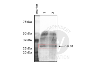
- Western Blotting
- Positivkontrolle
Adult mouse brain and cerebellum
- Negativkontrolle
- Bewertung
Passed. The CALB1 antibody ABIN6254097 works in WB at a 1:1000 dilution.
- Primärantikörper
- ABIN6254097
- Sekundärantikörper
goat anti-mouse IgG (H+L) HRP-conjugated (Thermo Fisher Scientific, G-21040)
- Full Protocol
- Homogenize tissues with cold lysis buffer containing 50 mM Tris HCl, 150 mM NaCl, 1% Triton X-100, 1 mM EDTA, and 1% protease inhibitor (Sigma P8340) using an ultrasonic homogenizer (MSE, SoniPrep 150) with 16 amplitude, 20 s on, 10 s off pulse for 60 s.
- Centrifuge tissue homogenates at 13,000 rpm for 20 min at 4 °C.
- Collect supernatants and determine total protein content using a Bradford assay.
- Denature 100 µg of total protein for 5 min at 90 °C and subsequently separate them on a denaturing 12% PAGE-SDS gel alongside a Precision Plus Protein Dual Color Standard (Bio-Rad, 160374).
- Electro-transfer proteins onto nitrocellulose membrane (Amersham Biosciences, RPN203D) ON in the cold room.
- Wash membrane 3x for 10 min with 0.01 M PBS containing 0.1% Tween-20 (PBST).
- Block membrane with PBST containing 2% bovine serum albumin for 1 h at RT.
- Incubate membrane with primary rabbit anti-CALB1 antibody (antibodies-online, ABIN6254097, lot GS116G) diluted 1:1,000 in PBST ON at 4 °C.
- Wash membrane 3x 10 min with PBST.
- Incubate membrane with secondary HRP-conjugated goat anti-mouse IgG (Thermo Fisher Scientific, G-21040) diluted 1:50,000 in PBST for 1 h at RT.
- Wash membrane 3x 10 min with PBST.
- Visualize proteins with SuperSignal West Atto Ultimate Sensitivity Substrate (Thermo Fisher Scientific, A38555) using a ChemiDoc Imaging System.
- Anmerkungen
Validierung #104530 (Western Blotting)![Erfolgreich validiert 'Independent Validation' Siegel]()
![Erfolgreich validiert 'Independent Validation' Siegel]() ValidierungsbilderProtokoll
ValidierungsbilderProtokoll -
- Buffer
- 100 μL in PBS + 50 % glycerol and 5 mM Sodium azide
- Konservierungsmittel
- Sodium azide
- Vorsichtsmaßnahmen
- This product contains Sodium azide: a POISONOUS AND HAZARDOUS SUBSTANCE which should be handled by trained staff only.
- Lagerung
- -20 °C
-
- Target
- CALB1 (Calbindin (CALB1))
- Andere Bezeichnung
- CALB1 (CALB1 Produkte)
- Molekulargewicht
- '28 kDa
- Gen-ID
- 793
- UniProt
- P05937
-


 (2 validations)


