RAF1 Antikörper (AA 31-130)
-
- Highlights
-
- High quality RAF1 Primärantikörper for the detection of RAF1.
- Reliable product with high standard validation data
- Target Alle RAF1 Antikörper anzeigen
- RAF1 (V-Raf-1 Murine Leukemia Viral Oncogene Homolog 1 (RAF1))
-
Bindungsspezifität
- AA 31-130
-
Reaktivität
- Human
-
Wirt
- Kaninchen
-
Klonalität
- Polyklonal
-
Konjugat
- Dieser RAF1 Antikörper ist unkonjugiert
-
Applikation
- ELISA, Immunofluorescence (Cultured Cells) (IF (cc)), Immunofluorescence (Paraffin-embedded Sections) (IF (p)), Immunohistochemistry (Paraffin-embedded Sections) (IHC (p)), Immunohistochemistry (Frozen Sections) (IHC (fro))
- Kreuzreaktivität
- Human
- Homologie
- Mouse,Rat,Dog,Cow,Horse,Rabbit
- Aufreinigung
- Purified by Protein A.
- Immunogen
- KLH conjugated synthetic peptide derived from human c Raf
- Isotyp
- IgG
- Produktspezifische Information
-
- What can the RAF1 Antibody ABIN733208 be used for?
- This polyclonal RAF1 Antibody detects RAF1. The RAF1 Antibody has been validated for various applications and can be used for the detection of RAF1 and derivatives by ELISA, Immunofluorescence (Cultured Cells), Immunofluorescence (Paraffin-embedded Sections), Immunohistochemistry (Paraffin-embedded Sections), Immunohistochemistry (Frozen Sections).
- What validation data is available for this RAF1 Antibody?
- The Primärantikörper is referenced in 1 publications and is characterized by a proven, very high reliability. It has currently 2 product images that show its performance in a variety of applications. The product is currently available in 100 μL quantity. RAF1 Antibody for the detection of RAF1 and derivatives.
- What is the function of RAF1?
- This gene is the cellular homolog of viral raf gene (v-raf). The encoded protein is a MAP kinase kinase kinase (MAP3K), which functions downstream of the Ras family of membrane associated GTPases to which it binds directly. Once activated, the cellular RAF1 protein can phosphorylate to activate the dual specificity protein kinases MEK1 and MEK2, which in turn phosphorylate to activate the serine/threonine specific protein kinases, ERK1 and ERK2. Activated ERKs are pleiotropic effectors of cell physiology and play an important role in the control of gene expression involved in the cell division cycle, apoptosis, cell differentiation and cell migration. Mutations in this gene are associated with Noonan syndrome 5 and LEOPARD syndrome 2. [provided by RefSeq, Jul 2008].
-
-
- Applikationshinweise
-
ELISA 1:500-1000
IHC-P 1:200-400
IHC-F 1:100-500
IF(IHC-P) 1:50-200
IF(IHC-F) 1:50-200
IF(ICC) 1:50-200 - Beschränkungen
- Nur für Forschungszwecke einsetzbar
-
- by
- Immunohistochemistry Core, NYU Langone
- No.
- #029651
- Datum
- 01.04.2014
- Antigen
- Chargennummer
- 130909
- Validierte Anwendung
- Immunohistochemistry
- Positivkontrolle
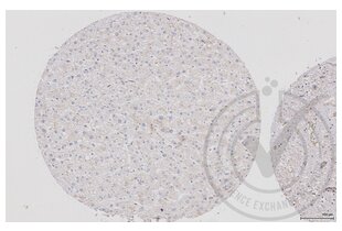
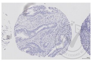
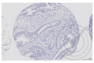
- Human colon villus epithelial cells
- Negativkontrolle
- Human liver
- Bewertung
- Raf1 exhibits uniformly low expression across all human tissues; higher expression found in colon, lower in liver. Tissue-specific expression is detected in colon, with expected low level expression in liver.
- Primärantikörper
- Antigen: V-Raf-1 Murine Leukemia Viral Oncogene Homolog 1 (RAF1)
- Catalog number: ABIN733208
- Lot number: 130909
- Sekundärantikörper
- Lot number: D07640BA
- Full Protocol
- Immunohistochemistry was performed on a Ventana NEXes automated platform; instrument manufacturer specific reagents are italicized.
- 1. Slides were preheated in convection oven at 60°C for 30 min
- 2. Deparaffinization procedure: - 3 changes of Xylene, 5 min each - 3 changes of 100% Ethanol, 3 min each - 3 changes of 95% Ethanol, 3 min each - Rinsed in distilled water, 3 changes
- 3. Heat retrieval procedure - Slides retrieved in 10.0 mM Citrate, pH6.0 in a 1000W microwave oven (~100°C) for 15 min. - Slides were allowed to cool (in citrate) for 30 min. - Slides were washed x 3 in Distilled water
- 4. NEXes instrument procedure, iView DAB paraffin protocol (*abridged*): - Slide chamber warmed to 37°C
- 5. Slides rinsed with *reaction buffer* x3
- 6. *iView Inhibitor (H2O2)* applied and incubated for 4 min
- 7. Slides rinsed with *reaction buffer*
- 8. Antibody Application - Primary antibody diluted 1:250 in PBS (100 microliter applied/slide) - Ventana Isotype control applied neat - Slides Incubated overnight at room temperature (~12 hours ~25°C)
- 9. Slides rinsed with *reaction buffer* x3
- 10. *iView Biotinylated IgG* applied and incubated for 8 min
- 11. Slides rinsed with *reaction buffer*
- 14. *iView Streptavidin-Horseradish Peroxidase* applied and incubated for 8 min
- 15. Slides rinsed with *reaction buffer*
- 16. *iView DAB/H2O2* applied and incubated for 8 min
- 17. Slides rinsed with *reaction buffer*
- 18. *iView Copper* applied and incubated for 4 min
- 19. Slides rinsed with *reaction buffer*
- 20. Slides washed in Dawn Detergent/tap water
- 21. Counterstain Procedure - Hematoxylin (Leica 560 MX) 30 sec - Slides washed in tap water, 1 min - Decolorized (10% Acetic Acid in 70% ethanol), 1 min - Slides washed in tap water, 1 min - Bluing (Austin Clear Ammonia), 1 min - Slides washed in tap water, 1 min
- 22. Dehydration/coverslipping procedure: - 3 changes of 95% Ethanol, 3 min each - 3 changes of 100% Ethanol, 3 min each - 3 changes of Xylene, 5 min each - Mounted with Permount
- 23. Imaging: Leica SCN 400F Whole Slide Scanner with Digital Image Hub and Leica Slidepath software
- Anmerkungen
- Deviations from protocol/procedure supplied by manufacturer:
- Step 1: Heated tissue 60°C for 30 minutes; manufacturer heats for 45 minutes.
- Step 2: No ethanol wash was performed during deparaffinization; manufacturer includes 1 wash of 80% ethanol for 3 minutes.
- Step 3.1: Slides were heated for 15 minutes; manufacturer provides a range of 15-20 minutes.
- Step 3.2: Slides were cooled for 30 minutes; manufacturer cools for 20 minutes.
- Step 4: Italicized reagents and incubation time are fixed instrument parameters.
- Step 5: Secondary species-specific serum block not used; manufacturer blocks with 5% normal goat serum for 2 hours.
- Step 8.1: Antibody diluted in PBS at 1:250; manufacture did not recommend diluent or dilution.
- Step 8.2.1: Primary antibody incubated at room temperature overnight; manufacturer incubates overnight 4°C with agitation.
Validierung #029651 (Immunohistochemistry)![Erfolgreich validiert 'Independent Validation' Siegel]()
![Erfolgreich validiert 'Independent Validation' Siegel]() ValidierungsbilderProtokoll
ValidierungsbilderProtokoll -
- Format
- Liquid
- Konzentration
- 1 μg/μL
- Buffer
- 0.01M TBS( pH 7.4) with 1 % BSA, 0.02 % Proclin300 and 50 % Glycerol.
- Konservierungsmittel
- ProClin
- Vorsichtsmaßnahmen
- This product contains ProClin: a POISONOUS AND HAZARDOUS SUBSTANCE, which should be handled by trained staff only.
- Lagerung
- 4 °C,-20 °C
- Informationen zur Lagerung
- Shipped at 4°C. Store at -20°C for one year. Avoid repeated freeze/thaw cycles.
- Haltbarkeit
- 12 months
-
-
: "Bone marrow mononuclear cell transplantation promotes therapeutic angiogenesis via upregulation of the VEGF-VEGFR2 signaling pathway in a rat model of vascular dementia." in: Behavioural brain research, Vol. 265, pp. 171-80, (2014) (PubMed).
-
: "Bone marrow mononuclear cell transplantation promotes therapeutic angiogenesis via upregulation of the VEGF-VEGFR2 signaling pathway in a rat model of vascular dementia." in: Behavioural brain research, Vol. 265, pp. 171-80, (2014) (PubMed).
-
- Target
- RAF1 (V-Raf-1 Murine Leukemia Viral Oncogene Homolog 1 (RAF1))
- Andere Bezeichnung
- c-Raf/Raf1 (RAF1 Produkte)
- Hintergrund
-
Synonyms: NS5, CRAF, Raf-1, c-Raf, CMD1NN, RAF proto-oncogene serine/threonine-protein kinase, Proto-oncogene c-RAF, RAF1, RAF
Background: Serine/threonine-protein kinase that acts as a regulatory link between the membrane-associated Ras GTPases and the MAPK/ERK cascade, and this critical regulatory link functions as a switch determining cell fate decisions including proliferation, differentiation, apoptosis, survival and oncogenic transformation. RAF1 activation initiates a mitogen-activated protein kinase (MAPK) cascade that comprises a sequential phosphorylation of the dual-specific MAPK kinases (MAP2K1/MEK1 and MAP2K2/MEK2) and the extracellular signal-regulated kinases (MAPK3/ERK1 and MAPK1/ERK2). The phosphorylated form of RAF1 (on residues Ser-338 and Ser-339, by PAK1) phosphorylates BAD/Bcl2-antagonist of cell death at 'Ser-75'. Phosphorylates adenylyl cyclases: ADCY2, ADCY5 and ADCY6, resulting in their activation. Phosphorylates PPP1R12A resulting in inhibition of the phosphatase activity. Phosphorylates TNNT2/cardiac muscle troponin T. Can promote NF-kB activation and inhibit signal transducers involved in motility (ROCK2), apoptosis (MAP3K5/ASK1 and STK3/MST2), proliferation and angiogenesis (RB1). Can protect cells from apoptosis also by translocating to the mitochondria where it binds BCL2 and displaces BAD/Bcl2-antagonist of cell death. Regulates Rho signaling and migration, and is required for normal wound healing. Plays a role in the oncogenic transformation of epithelial cells via repression of the TJ protein, occludin (OCLN) by inducing the up-regulation of a transcriptional repressor SNAI2/SLUG, which induces down-regulation of OCLN. Restricts caspase activation in response to selected stimuli, notably Fas stimulation, pathogen-mediated macrophage apoptosis, and erythroid differentiation.
- Gen-ID
- 5894
- UniProt
- P04049
- Pathways
- MAPK Signalweg, RTK Signalweg, Fc-epsilon Rezeptor Signalübertragung, Neurotrophin Signalübertragung, cAMP Metabolic Process, Stem Cell Maintenance, Hepatitis C, Autophagie, Signaling of Hepatocyte Growth Factor Receptor, VEGF Signaling, BCR Signaling
-


 (1 reference)
 (1 Validierung)


